
Histopathology and Anatomy Laboratory
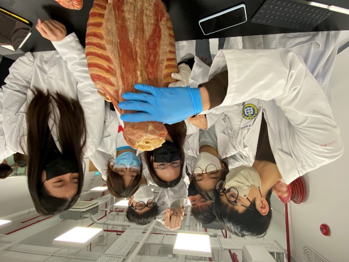
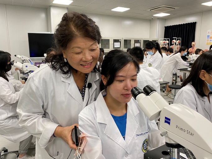
The Histopathology and Anatomy Laboratory is equipped with teaching microscopes, light microscopes, anatomical mannequins and specimens, as well as the world’s most advanced Anatomage Table 3D interactive virtual human anatomical dissection instrument, which is used by hundreds of world-leading universities and hospital units globally, such as Harvard University, Stanford University, University of Pennsylvania, University of Miami, and University of Iowa.
This allows medical students to conduct a new digital interactive anatomy teaching course. 1:1 real life size, 3D image software, multi-touch screen, image-guided surgical device and high-tech system with built-in real human anatomy model to present the human gross anatomy images in real life. It is particularly suitable for radiology classes related to imaging, radiation, surgical cases review, patient counseling and anatomy education. This allows students to explore, dissect and understand various parts of the human body. This device can store more than 1,000 real pathological 0.2 mm slices. The exclusive technology of Anatomage can clearly present the structure of microvessels and nerves. There are 1,500 clinical cases in its diverse database, including brain tumours, cancers, ectopic pregnancy, conjoined twins, etc. The built-in data is directly imported from CT, MRI (no retouching adjustment) and is shown in UHQ ultra-clear images.
The device is also equipped with an anatomical data collection system that allows students to view a wide range of CT and MRI medical images and scans for anatomy and physiology courses, including: cross-sectional anatomy, X-ray, CT or MRI slides, images of disease or abnormality, anatomical variations, bone fractures, or structural identifications of cardiovascular conditions or disorders (along with pathological cases).


