|
Imaging core facility
Visual evidence is crucial in biomedical discovery - seeing is believing. The preparation and visualization of tissue sections from animal models or human patient samples is a crucial step in translating laboratory research into clinically relevant insights. This core facility is designed to support researchers in every aspect of tissue-based studies, from histological preparation to advanced imaging and flow cytometry. With cutting-edge equipment and expert technical support, the imaging core is a vital resource for researchers working on disease mechanisms, therapeutic interventions, and regenerative medicine.
Our facility provides high-quality histology services alongside a wide range of imaging and flow cytometry technologies. These tools allow researchers to gain a deeper understanding of tissue architecture, cellular behavior, and molecular interactions, driving translational research that bridges the gap between the bench and the clinic.
Capabilities and Equipment
Advanced Imaging and Analysis Technologies
Our core is equipped with state-of-the-art imaging systems, enabling detailed exploration of cellular structures and molecular interactions within tissues. Key imaging modalities available include:
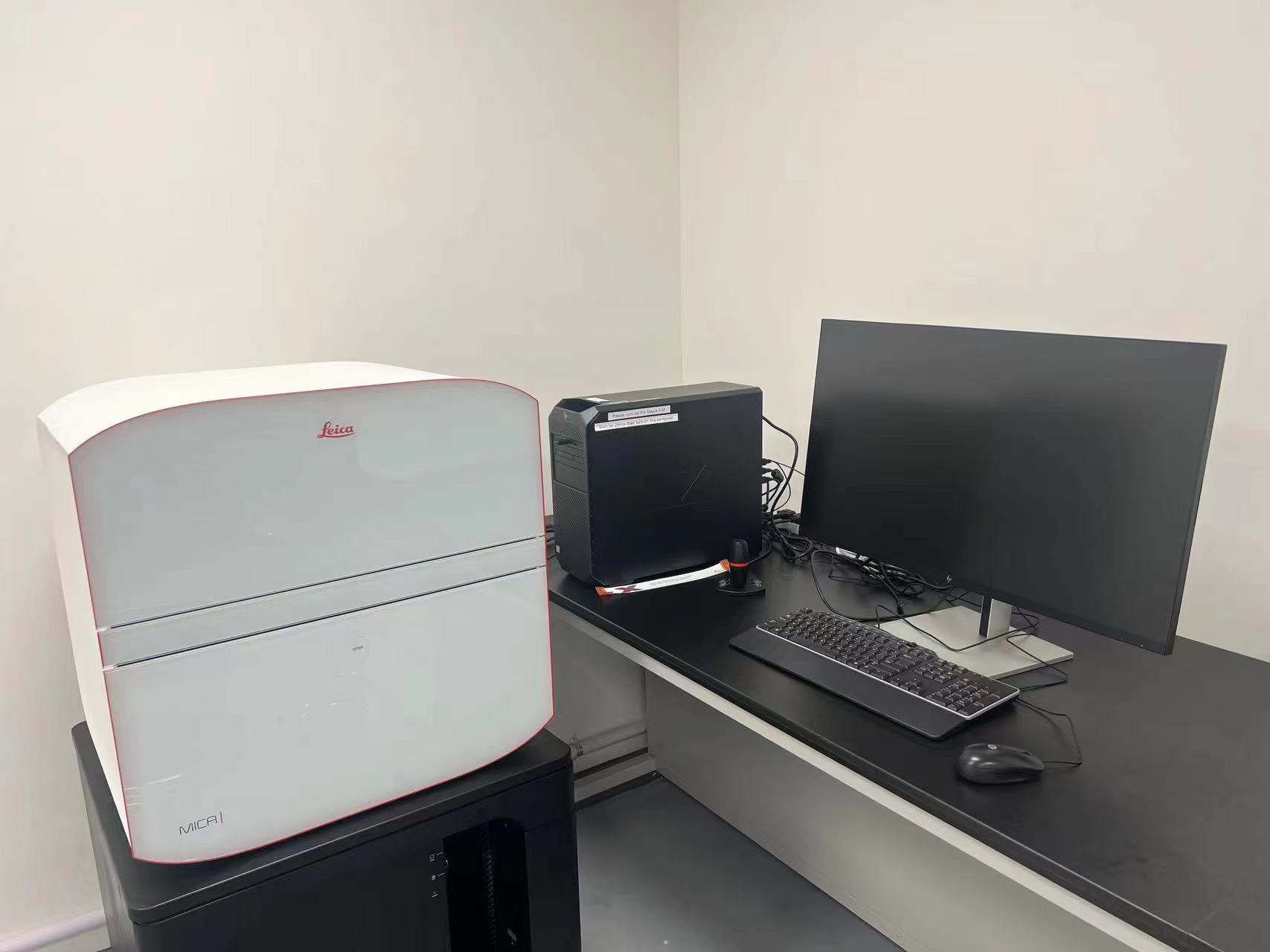
The Leica MICA Confocal Microscope
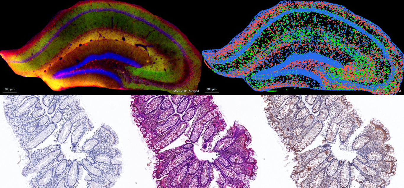
Confocal microscopy provides high-resolution imaging of tissue sections and living cells, allowing researchers to capture optical sections and reconstruct 3D images. The Leica MICA Confocal Microscope supports multi-channel fluorescence imaging using FluoSync technology for unmixing multicolor images. The MICA system provides exceptional clarity for studies involving protein localization, cell signaling, and dynamic cellular processes.
Leica AIVIA AI-assisted Advanced Image Analysis Suite
The Leica AIVIA AI-assisted Advanced Image Analysis Suite provides powerful tools for automated and semi-automated analysis. Aivia combines powerful artificial intelligence-guided image analysis and visualization solutions for researchers to extract meaningful data from their images. The AI-enabled tools in Aivia, coupled with deep learning to enhance, segment and predict signals in your images provides new means of analysis and new interpretation of data.
EVOS M5000 Imaging System
The EVOS M5000 Imaging System provides a rapid and simple way to image samples in 4-colour fluorescence or transmitted light. The imaging system comes with an onstage incubator, which allows for live-cell analysis with control of temperature, humidity and gases. The Celleste software is simple and easy to use, making the EVOS M5000 Imaging System ideal expeditive visualization of life and fixed samples.
Flow Cytometry Services

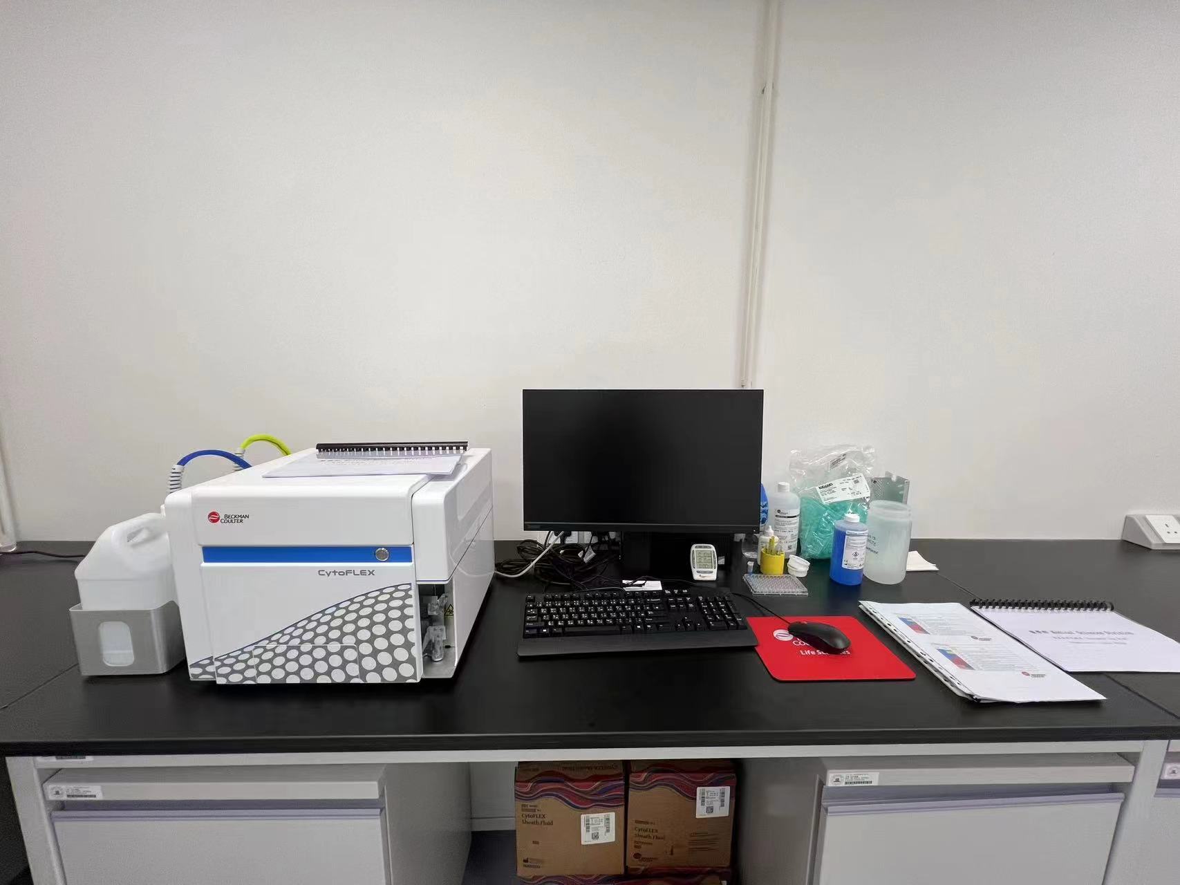
Flow cytometry is a key technique for analyzing the physical and chemical characteristics of cells or particles in suspension. Our facility is equipped with the Beckman Coulter CytoFlex Flow Cytometry System, which offers multi-parameter analysis, enabling researchers to measure a wide range of cell properties, such as size, granularity, and fluorescence intensity. This system is ideal for immunophenotyping, cell cycle analysis and apoptosis for downstream applications.
Whole-Slide Imaging
For researchers requiring high-resolution scanning of entire tissue sections, our facility offers the Zeiss Axioscan System, which provides automated, high-speed slide scanning. This system generates digital images of entire slides, allowing for detailed analysis and easy sharing of data with collaborators. Whole-slide imaging is particularly useful for pathological studies, enabling quantitative analysis of large tissue areas and high-throughput screening of samples.


